Central Anatomy and Physiology
(including the initial peripheral retrocochlear system)
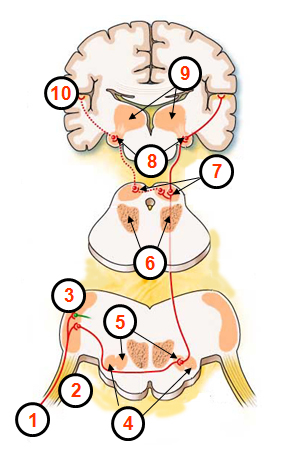 Brain Schematic
1. Type I Neurons
2. Cochlear Nerve
3. Cochlear Nucleus
4. Lateral Superior Olives
5. Medial Superior Olives
6. Reticular Formation
7. Inferior Colliculus
8. Medial Geniculate Body
9. Thalamus
10. Auditory Cortex
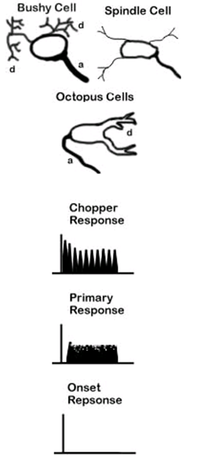 Central System Intensity Coding Cells
• Bushy Cells – Primary Response
These cells have responses very similar to those in the auditory nerve. The primary difference is that spontaneous activity is decreased by stimulation by adjacent frequencies. This leads to an even sharper tuning curve than seen in auditory nerve cells.
• Spindle Cells – Chopper Response
Spindle cells produce a “chopper” response. This can be best described as a rapid consistent response at the beginning of the stimulus followed by a train of spikes with a specific between spike interval for the duration of the stimulus.
• Octopus Cells – Onset Response
These cells respond only at the onset of a specific frequency or frequency range at higher amplitudes
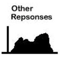
There are also other cells with more complex responses.
Cochlear Nucleus
CN is Almost like the switchboard of the central auditory pathway. As many as 20 different morphologic cells have been identified – few are understood
- Anterior Ventral Cochlear Nucleus (AVCN)
- Posterior Ventral Cochlear Nucleus (PVCN)
- Dorsal Cochlear Nucleus (DCN)
Anterior Ventral Cochlear Nucleus (AVCN)
• The entryway to the “Binaural Pathway“
• Through the binaural pathway, the AVCN provides input to the Superior Olivary Complex (SOC). This pathway is likely to be involved in parts of spatial localization and other tasks requiring convergence of information from both ears, but it’s not totally clear how.
• Bushy and Spindle Cells
Posterior Ventral Cochlear Nucleus (PVCN)
• The PVCN is the starting point of the “Intermediate Brainstem Pathway.”
• The functionality of this pathway is poorly understood.
• Connects to the Periolivary Nuclei (PON) and ascends up the Lateral Lemniscus (LL) where it connects to nuclei of the LL and Inferior Colliculus (IC).
• Bushy, Spindle, and Octopus
Dorsal Cochlear Nucleus (DCN)
• “The Monaural Contralateral Pathway”
• carries information from only the contralateral ear
• Bushy, Spindle, Octopus, and Others
Superior Olivary Complex (SOC)
• Medial Superior Olivary Nucleus (MSON)
• Lateral Superior Olivary Nucleus (LSON)
• Medial Nucleus of the Trapezoid Body (MNTB)
• Periolivary Nuclei (PON)
• The SOC processes information about interaural delays and amplitudes (localization).
Cell types in the SOC
The first letter describes the contralateral response, either E (excitatory) or I (inhibitory.) The second letter similarly describes the neuron’s response to sound in the ipsilateral ear.
• EE – excited by both contralateral and ipsilateral
• EI – excited by contralateral, but inhibited by ipsilateral
• IE – excited by ipsilateral, but inhibited by contralateral
• II – inhibited by both contralateral and ipsilateral
Medial Superior Olivary Nucleus (MSON)
• MSON neurons respond to both binaural and monaural stimuli, however they respond more to binaural sounds
• The response tend to be frequency selective and the best frequency is typically the same for both ipsilateral and contralateral input
• The characteristic frequencies in the MSON tend to be in the low end of the hearing spectrum.
• Neurons in the MSON respond best to a specific delay between ipsilateral and contralateral stimulus termed the “characteristic delay.” They also have a “least characteristic delay” to which they respond worse than to monaural input.
• Mix of cell types
• Efferents to outer hair cells – primarily inhibitive
Lateral Superior Olivary Nucleus (LSON)
• Neurons in the LSON are sensitive to intensity differences between sounds in the ipsilateral and the contralateral ears.
• Almost all IE cells
• These best frequencies are predominantly in the high frequency range complimenting the range of frequencies over which the MSON is responsive.
• Efferents to inner hair cells
Medial Nucleus of the Trapezoid Body (MNTB)
• The MNTB is responsive exclusively to sound presented to the ear contralateral to the nucleus itself as a function of level.
• Each cell responds best to a characteristic frequency. Two response types are found in the MNTB: a chopper type similar to spindle cells in the AVCN and a primary type which is very similar to those of bushy cells in the AVCN.
Periolivary nuclei (PON)
• The entire superior olive complex is surrounded by small cellular groups known as the preolivary or periolivary nuclei.
• The function is poorly understood
Lateral Lemniscus
The LL is primarily a tract of axons ascending the brainstem from other structures passing through the LL nucleus cells. The LL cells are sensitive to changes in both the timing and the amplitude of sound.
• Dorsal
• Ventral
• Intermediate
Dorsal Nucleus of the Lateral Lemniscus (DNLL)
• The cells of the DNLL appear to respond best to binaural inputs. Onset and complexly tuned sustained responses have been recorded, but very little is really known
Ventral Nucleus of the Lateral Lemniscus (VNLL)
• Very little spontaneous activity is present in the VNLL.
• When elicited, responses in the VNLL are strongest to sound in the contralateral ear. The tuning curves of these cells are very broad and moderately complex.
• The VNLL is the most likely region of the NLL to be involved in the startle reflex.
Intermediate Nucleus of the Lateral Lemniscus (INLL)
• Cells in the INLL show little spontaneous activity and have broad tuning curves.
• Chopper, onset-like, and sustained responses have all been found in the INLL, but few models have been developed relating the inputs of these cells to their responses.
Inferior Colliculus (IC)
• Information integration
• Switchboard to higher auditory processes
• Provides a spatio-topic map of the auditory environment. Sends information to the Superior Colliculus, where saccadic (pronounced: sa-cod-ic) eye movement (quick, simultaneous movements of both eyes in the same direction) is generated
• May also provide a modulo-topic representation (frequency and amplitude changes). Possibly the beginning of speech phoneme identification
Thalamus
• The thalamus is the last relay site on the way to the cortex for almost all sensory information, including auditory, visual, and somatosensory (body sense – as opposed to 5 senses). It is shaped like a football and has been functionally and morphologically divided into a large number of nuclei. The primary nucleus involved in the auditory pathway is the Medial Geniculate Body (MGB), but several other nuclei are at least partially responsive to auditory stimulation. The MGB has itself been divided into three subneclei based on functional as well as morphological distinctions. These divisions are Ventral (VMGB), Medial (MMGB), and Dorsal (DMGB).
Ventral Subnucleus of the Medial Geniculate Body (VMGB)
• The VMGB is thought to be primarily responsible for relaying frequency, intensity and binaural information to the cortex.
• Spatiotopic and modulotopic maps (as in the IC) however have not been well supported by mammalian studies (differences between humans and other animals?).
• Both monaural (10%) and binaural cells (90%) exist in the MGB. The monaural cells are primarily responsive to contralateral sound. Binaural cells are typically EE or EI types.
Medial Subnucleus of the Medial Geniculate Body (MMGB)
• The MMGB seems to functionally be responsible for detection of the relative intensity and duration of a sound.
• Binaural interactions found in the MMGB include EE, EI, and IE types. Both broadly and narrowly tuned cells have been observed. A type of intensity tuning has also been observed. In this type of cell, the response actually decreases as sound intensity increases above a specific level.
• Almost all cells in the MMGB appear to respond for the duration of the stimulus, and have very little adaptation. Individual cells often have more than one CF and are broadly tuned.
• Anesthetics tend to have large effects on cells within the MMGB, making responses difficult to study.
• MMGB cell-behavior is complicated by the fact that sensory stimulation from other modalities modifies the responsiveness of many, but not all, cells in the MMGB.
Dorsal Subnucleus of the Medial Geniculate Body (DMGB)
• Many types of responses are present in the DMGB which appear to vary across region.
• Generally, the responses are broadly tuned, but some cells appear to respond only to complex stimuli.
• Other cells are multi modal, often responding to somatosensory as well as auditory stimuli.
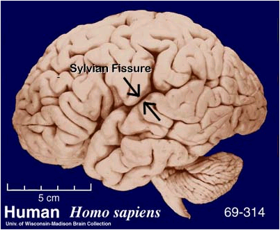 Auditory Cortex
• In the Lateral Sulcus (Sylvian Fissure) of the Temporal Lobe (part of the telencephalon – cerebrum)
• There does appear to be a tonotopic organization in auditory cortex with higher frequencies located more medially.
• It is likely that there are several tonotopic maps represented in the cortex, each with specific functions in demodulation of speech and other sounds.
• There also appears to be a spatiotopic map with sounds from the contralateral hemifield being more excitatory in a given hemisphere.
• Regions with specific temporally sensitive responses have also been found which may play a role in several phenomena of perception including virtual pitch perception, timbre discrimination, spatial localization, or even noise filtering.
Anatomy of the Brain
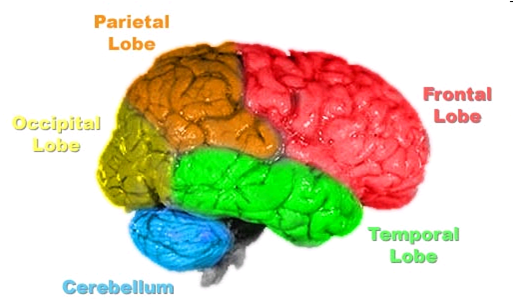
Lateral Sulcus divides the frontal lobe and parietal lobe above
from the temporal lobe below
(image: http://upload.wikimedia.org/wikipedia/en/5/5b/Brain-anatomy.jpg)
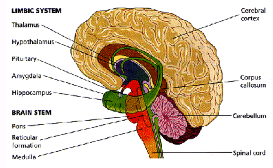
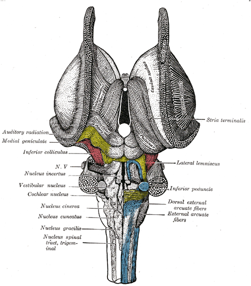
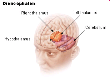
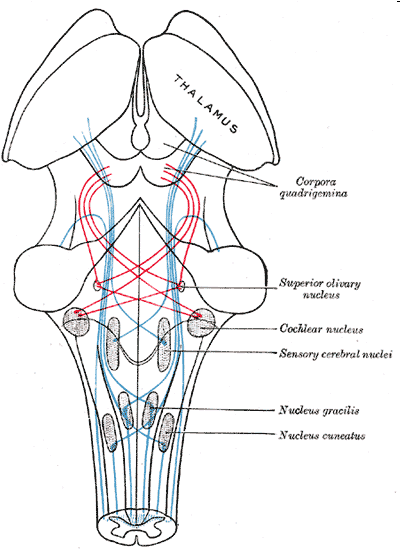
Image: http://upload.wikimedia.org/wikipedia/commons/5/52/Gray691.png
Others: Source Unknown